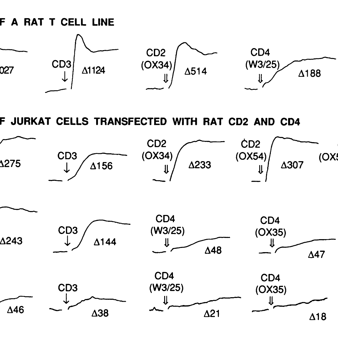
We were among the first groups to consider the problem of how recognition at the cell surface can be both weak and specific, and perhaps the first to emphasize its novel features.
To understand how cells interact it’s helpful to know in detail the structures and interactions of the proteins involved. Our success in this work depended entirely on our ability to make first-class soluble T-cell surface proteins. For this we have to thank having access to the glutamine synthetase-based gene expression system, and expert training in the use of the system by Prof Neil Barclay at what was then the MRC Cellular Immunology Unit.
To understand how cells interact it’s helpful to know the structures and interactions of the proteins involved. SJD went to Oxford initially to help determine the structure of the AIDS virus receptor, CD4. Alan Williams had argued that because protein crystallization was unpredictable several species should be tried in case human CD4 was especially difficult. We started work on rat CD4 and published the first (problematic) crystals, but the first structure to be solved comprised the N-terminal two domains of human CD4 by the groups of Steve Harrison and Wayne Hendrickson. The work on multiple species was vindicated; it just hadn’t worked in our favour.
We had tried the N-terminal two domains of rat CD4 also, and a crucial difference was that the rat protein carried a domain 2 N-glycan and it wouldn’t crystallize. This prompted us to try to solve the “glycosylation problem”, i.e. the need to deglycosylate glycoproteins of crystallographic interest. Glycosylation is an issue for crystallization studies because “normal” N-glycans are large, flexible and extremely heterogeneous and therefore work against the formation of reproducible lattice contacts in crystals. We chose the general approach of deglycosylating the protein of interest after we had purified it (1) because co-translational glycosylation is often required in order for proteins to fold correctly in mammalian cells in the first instance and because we had made a large commitment to mammalian expression (2).
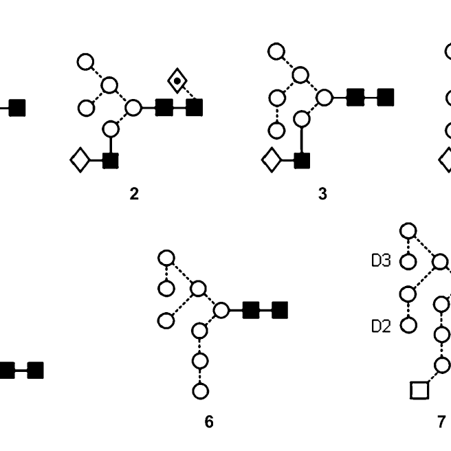
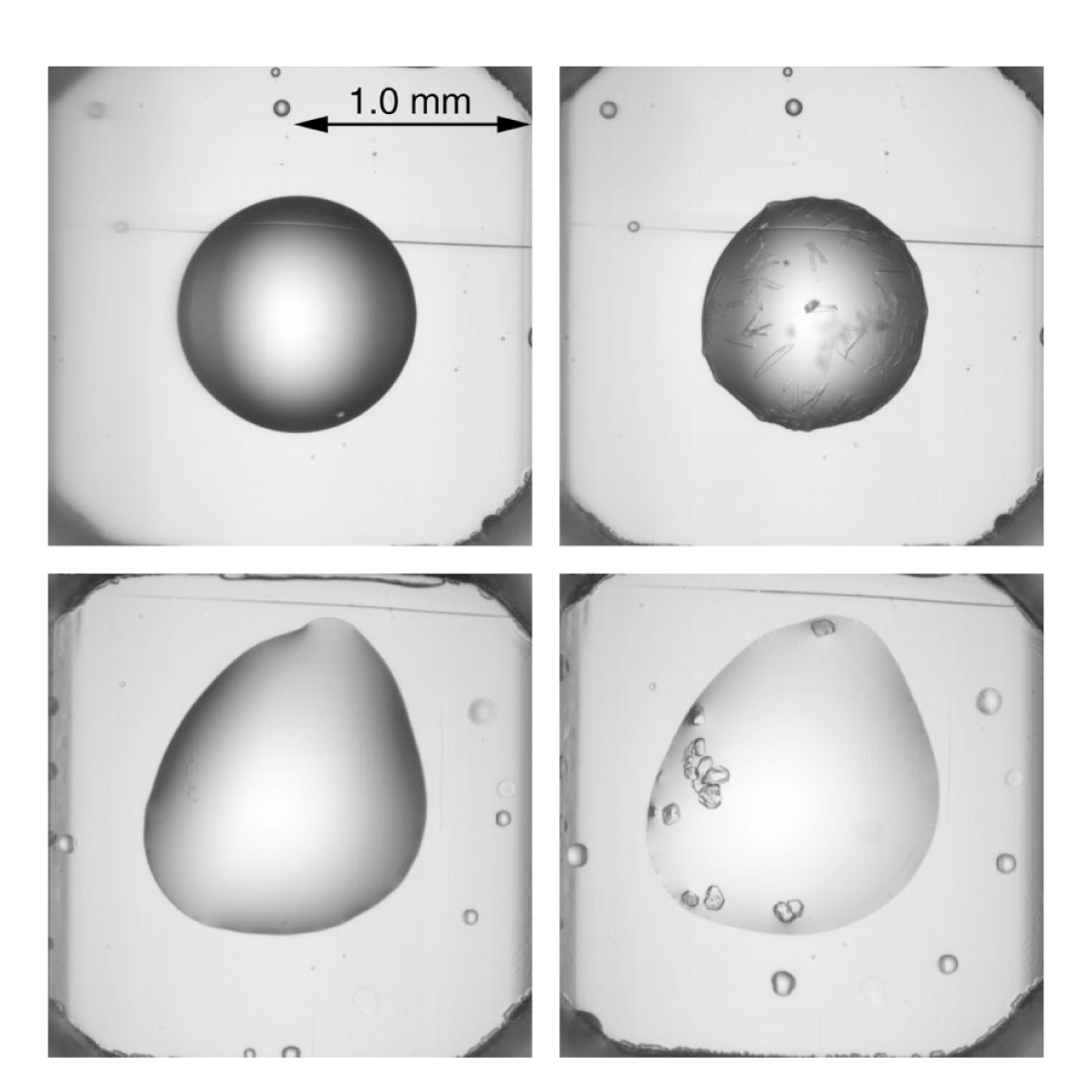
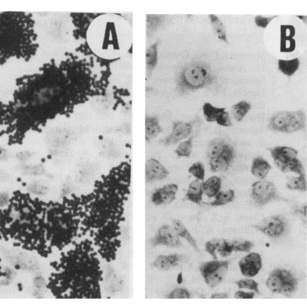
We generated deglycosylatable forms of glycoproteins by arresting the processing of the N-glycans at an endoglycosidase (Endo) H- or Endo F1-sensitive, largely oligomannose stage using lectin-resistant Chinese hamster ovary (CHO)-derived cell lines deficient (most importantly) in UDP-N-acetyl-d-glucosamine:α-3-d-mannosidase β1,2-N-acetylglucosaminyltransferase I (e.g. Lec3.2.8.1 cells), or by adding the α-glucosidase I inhibitor N-butyldeoxynojirimycin to CHO cell cultures expressing our proteins. This idea emerged after we learnt that one of the chief (perhaps only) advantages of insect-cell expression is that insect cells add fairly simple, relatively easily removed N-glycans to proteins. The first protein for which the new mammalian cell-based method was tried was rat CD2 (four N-glycans distributed over two domains), which produced beautiful crystals in the first hanging-drop we set up after expressing the protein in Lec3.2.8.1 cells and digesting it with Endo H (1) (Fig. 1).
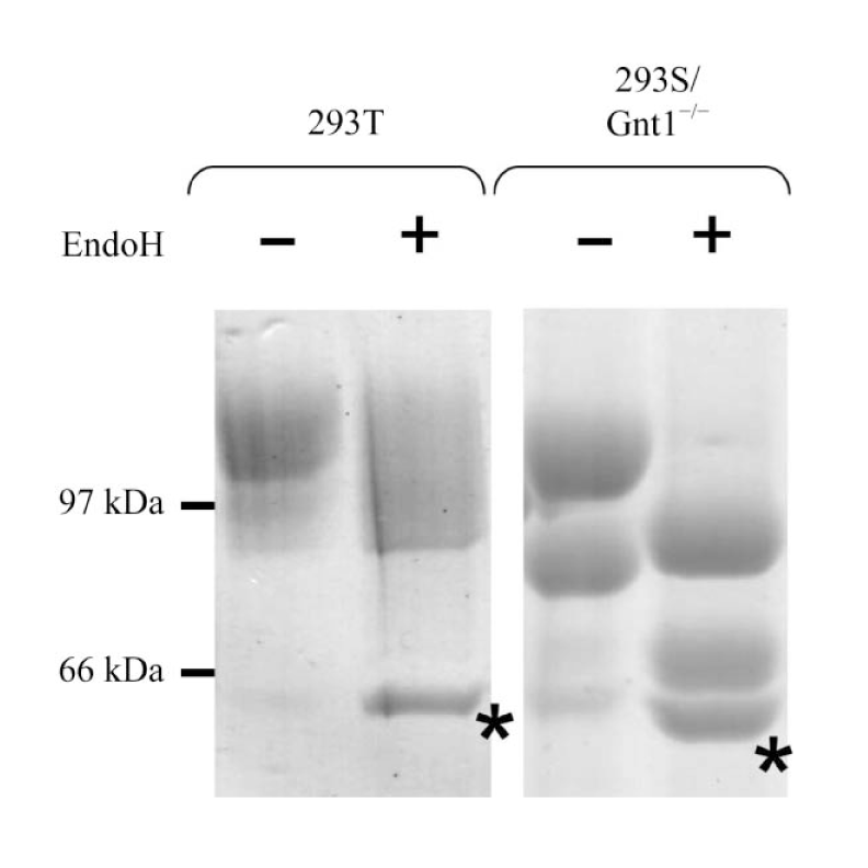
We have found that the polydispersity in solution of most Endo H/F1 treated proteins is indistinguishable from that of wild-type proteins, whereas glycoproteins completely deglycosylated with, for example, PNGase F, tend to aggregate. We subsequently developed tools (3) for producing deglycosylatable proteins in transient mammalian expression systems based on human embryonic kidney 293T cells, using the alkaloids kifunensine and swainsonine, which are inhibitors of α-mannosidases I and II, respectively. This coincided with great improvements in the yields, efficiency and scalability of transient expression protocols owing to the advent of new episomal expression vectors, transfection protocols and the introduction of a cheap transfection reagent, polyethylenimine, together with the development of multiplex, deep-well tissue culture methods. These types of methods are now widely used in glycoprotein crystallography.
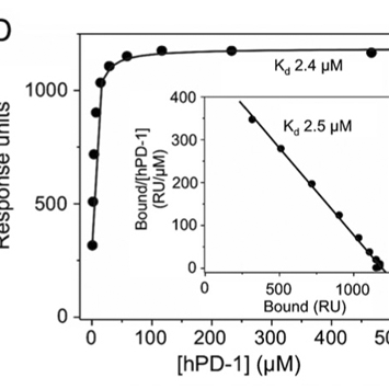
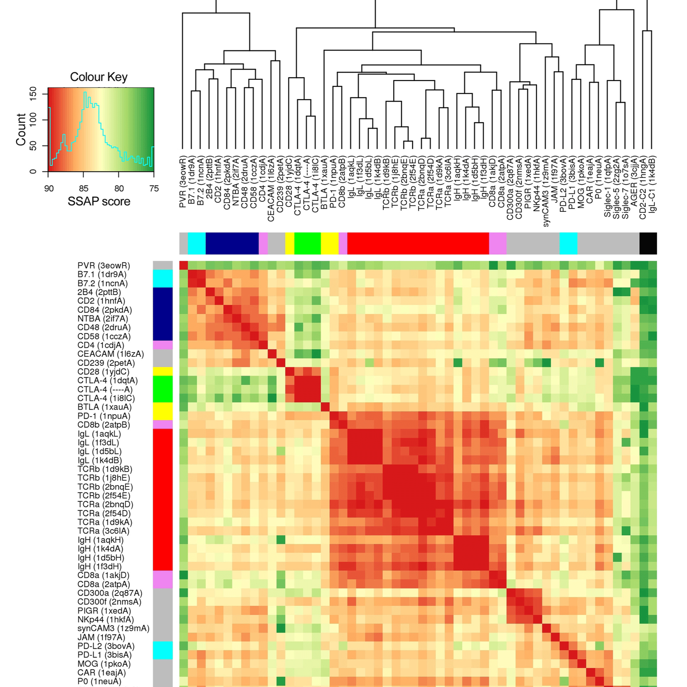
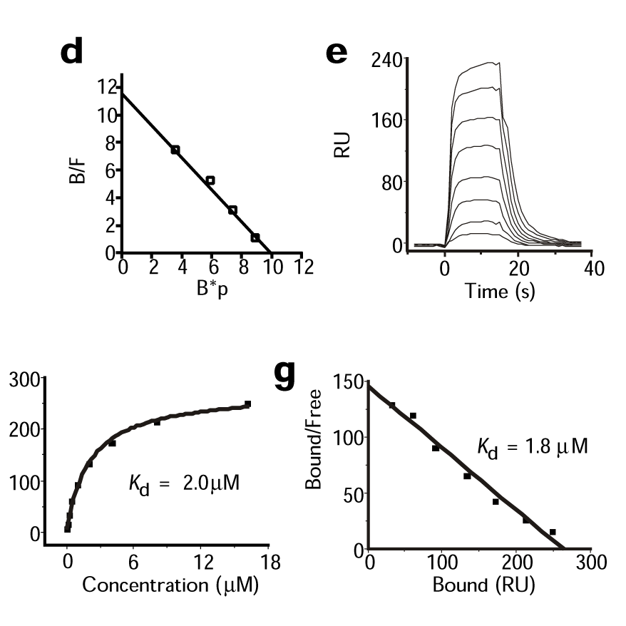
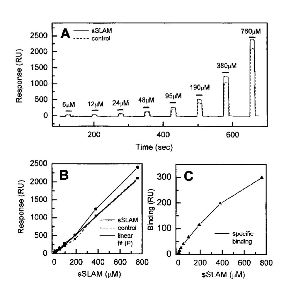
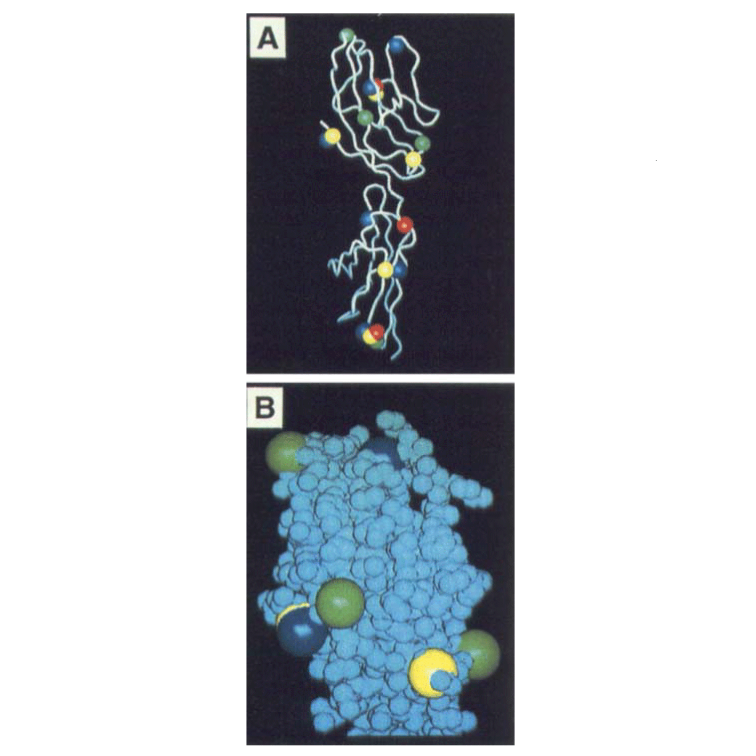
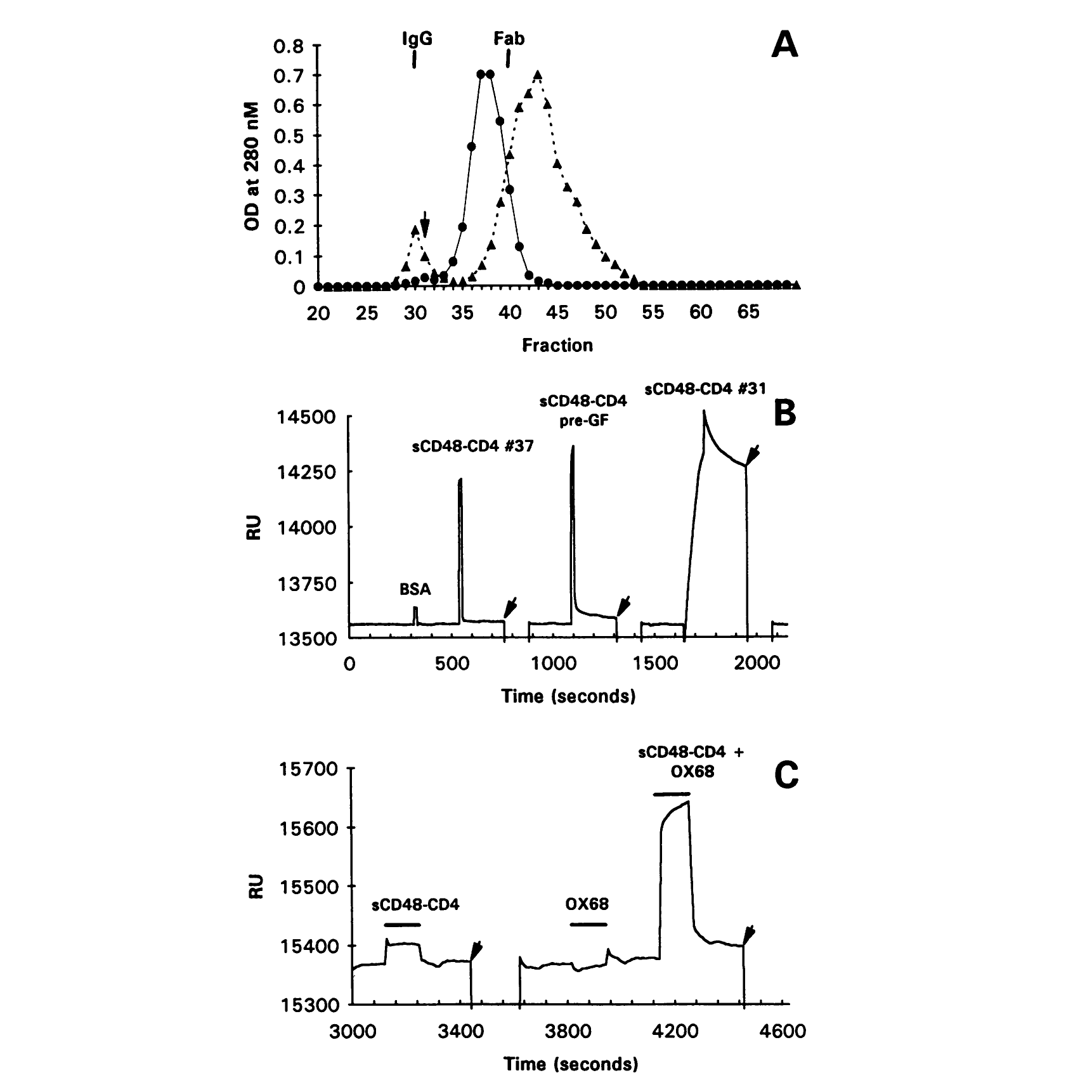
Access to the structures and to surface plasmon resonance-based assay (Biacore™) technology then allowed us to get at the problem of how low-affinity, high-specificity recognition is achieved at the cell surface. Our early work on CD2 based on the rat CD2 structure highlighted the importance of charged residues (4) (Fig. 2). We then determined the crystal structure of the ligand-binding domain of the CD2 ligand, CD48, and characterized its interactions with CD2 and 2B4. The relatively simple arrangement of charged residues on the flat binding face of CD48 seems ideally suited to binding multiple ligands, while maintaining low affinity for each of them. We speculated that cross-reactive interactions of this type fuelled the initial expansion of CD2-related genes. Overall, the data suggest that the evolutionary diversification of interacting cell surface proteins is rapid, and constrained only by the requirement that binding is weak and specific.